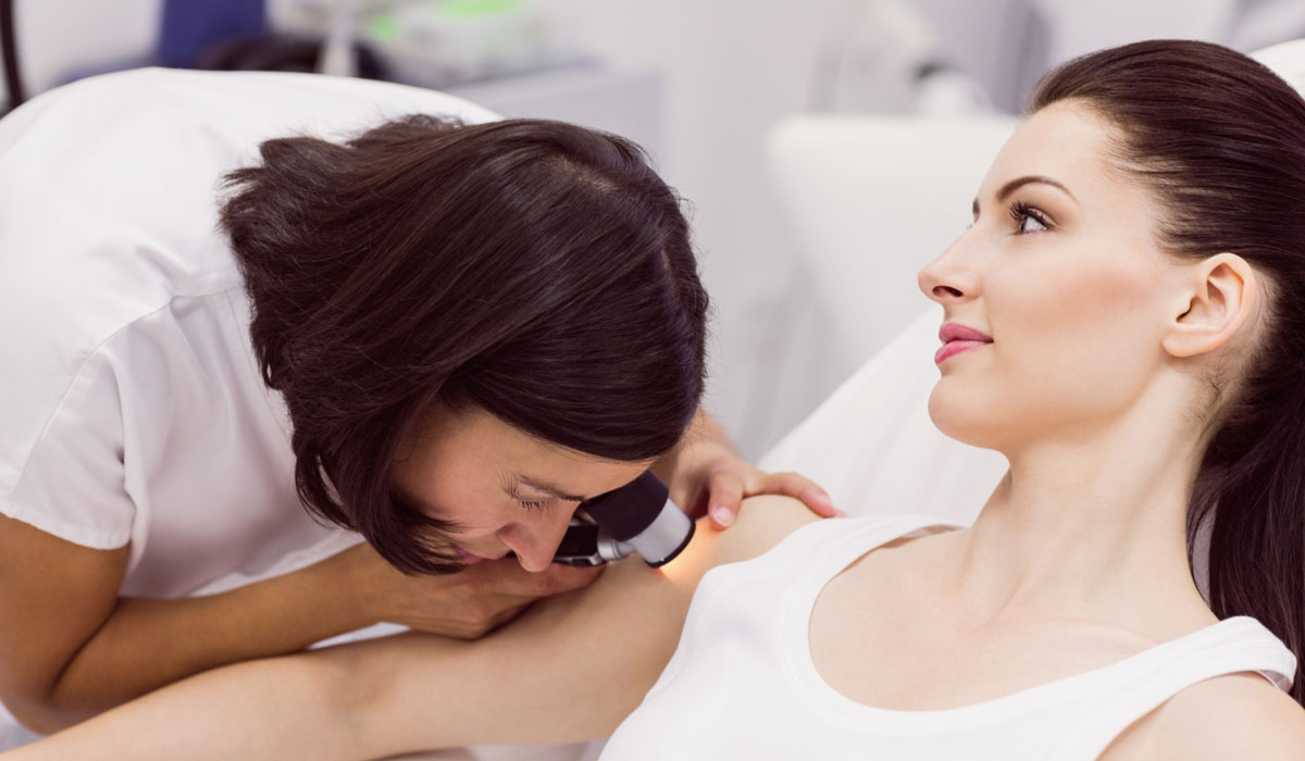
A skin check is a comprehensive evaluation of your skin health conducted by a trained healthcare provider to detect skin cancer and other skin conditions early. It involves visual inspection, discussion of medical history and risk factors, focused examination of specific lesions, and patient education regarding skin health and preventive measures.


During a skin check, a dermatologist or healthcare provider performs a thorough examination of your skin to assess for any abnormalities, including signs of skin cancer or other skin conditions. Here’s what typically happens during a skin check:
Visual Examination
The dermatologist will visually inspect your skin from head to toe, examining areas that are commonly exposed to the sun as well as those that are typically covered. They use a bright light and possibly a magnifying glass (dermatoscope) to get a closer look at moles, spots, and other skin lesions.
Key Points:
- Comprehensive Assessment: The dermatologist examines your entire body, including areas that may be difficult for you to see on your own.
- Focused Inspection: They focus on identifying any suspicious growths, changes in moles, or new lesions.
- Documentation: Any concerning findings are documented for future reference and comparison.
Discussion and History Taking
The dermatologist will ask about your medical history, including any personal or family history of skin cancer, previous skin conditions, sun exposure habits, and any concerns you may have regarding your skin.
Key Points:
- Risk Assessment: They assess your risk factors for skin cancer based on personal and family history, skin type, and sun exposure.
- Patient Education: They provide information about skin cancer risk reduction, sun protection measures, and the importance of self-examination.
Examination of Specific Lesions
If any suspicious lesions are identified during the visual examination, the dermatologist may perform a closer evaluation using a dermatoscope or may recommend a biopsy to obtain a tissue sample for further analysis.
Key Points:
- Dermatoscopy: A dermatoscope (hand-held device with magnification and light) helps in examining moles and lesions in more detail.
- Biopsy: If a lesion appears concerning, a biopsy involves removing a small sample of tissue for laboratory examination to determine if cancerous cells are present.
Education and Recommendations
Based on the findings of the skin check, the dermatologist will discuss their observations with you and provide recommendations for further evaluation, monitoring, or treatment if necessary.
Key Points:
- Follow-up Plan: They may recommend regular self-examinations and follow-up visits for monitoring specific lesions or for high-risk individuals.
- Treatment Options: If skin cancer or other skin conditions are detected, they discuss treatment options and potential outcomes.

